
Abstract
Background: Enamel pearls are developmental anomalies consisting of ectopic enamel, with or without dentine and pulp tissue. They most commonly occur on maxillary second and third molars and are less frequently associated with mandibular third molars. Although they may have periodontal implications, their anatomical location can also influence surgical management. Case report: A 32-year-old female was referred with recurrent episodes of pain and inflammation associated with the mandibular third molars. Both lower third molars were partially erupted and mesioangular, with no active pericoronitis or periodontal pathology at presentation. An orthopantomogram demonstrated an intimate relationship between both mandibular third molars and the inferior dental canals. Subsequent cone beam computed tomography (CBCT) demonstrated three convergent roots associated with the mandibular right third molar and an enamel pearl arising from the mesial root and extending lingually around the middle third of the root. The inferior dental canal demonstrated pronounced narrowing and deviation around the tooth, with the enamel pearl extending into the canal and towards the lingual cortical plate. The mandibular left third molar also demonstrated an intimate relationship with the inferior dental canal. Management and outcome: The increased risk of inferior alveolar nerve injury associated with surgical removal was discussed alongside the options of observation, coronectomy and complete extraction. Following informed discussion, the patient elected to monitor the mandibular third molars. The carious maxillary third molars were subsequently removed under local anaesthesia. Conclusion: This case highlights an unusual enamel pearl associated with a mandibular third molar and its potential implications for third molar surgery. CBCT provided important three-dimensional information regarding the relationship between the enamel pearl, tooth roots and inferior dental canal, facilitating risk assessment, treatment planning and informed patient decision-making.
1. Introduction
Enamel pearls are extensions of tooth structure, either enamel, or having more complex features like enamel dentine, or enamel dentine pulp tissues. The formation of these is generally believed to arise from remnants of Hertwigs root sheath (HERS), which can develop ectopic enamel from misplaced ameloblasts of HERS. They typically form on maxillary second or third molars. This case depicts an enamel pearl associated with the root surface of a mandibular third molar and the potential consequences in relation to further third molar surgery.
2. Background
A 32-year-old female patient attended an outpatient clinic appointment following a referral from their general dental practitioner. This was due to repeated episodes of discomfort surrounding the lower third molars and concerns of detrimental effects to the lower second molars. On attendance the patient reported that occasionally they had symptoms of pain and inflammation localised around the gingival tissues of the lower third molars. They reported that the incidents settled without intervention and this has happened multiple times over the past year. There was no history of spreading facial swelling during the consultation and no episodes of pericoronitis requiring antibiotics to treat.
On examination the patient had no extraoral pathology. Intraorally both upper third molars had caries into the inner third of dentine however were not tender to percussion. Both lower third molars were partially erupted and mesially angulated. There was no clinically detectible inflammation surrounding the lower third molars on assessment and no increased pocketing around these teeth. There was no clinical evidence of caries on the lower second molars or on the occlusal surface of both lower third molars. Oral hygiene was excellent and the remaining dentition was sound and unrestored. The soft tissues within the oral cavity were free of pathology.
An initial Dental Panoramic Radiograph (Figure 1) was taken to assess the dentition for caries, and the lower wisdom teeth for angulation, pathology and proximity to the inferior dental canal. This OPT suggested there was an intimate relationship with banding bilaterally on the third molars where the inferior dental canal overlaps the teeth (Figure 1). On the OPT it was also evident that the upper right and left third molars also had caries present in both teeth however the remaining dentition was sound.
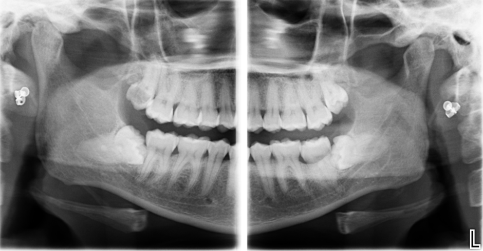
This prompted a request for a cone beam CT scan of bilateral lower third molars to assess the proximity of the inferior dental canal and the roots of the third molar. This was required to assess the risk of inferior dental nerve damage during the surgical removal of these lower third molars.
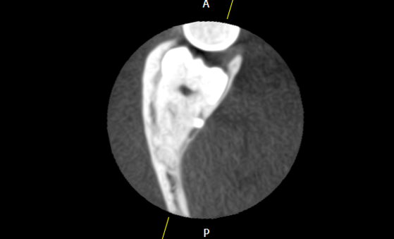
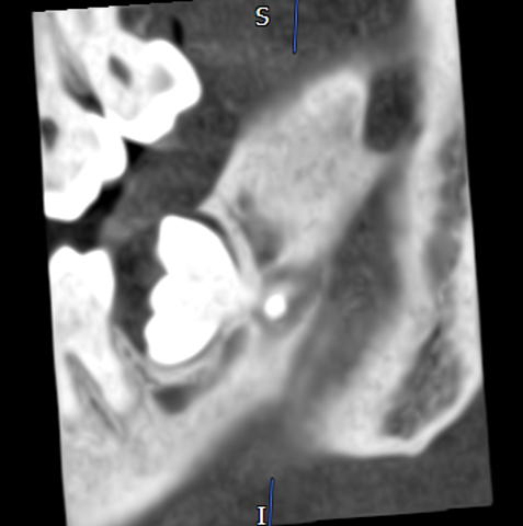
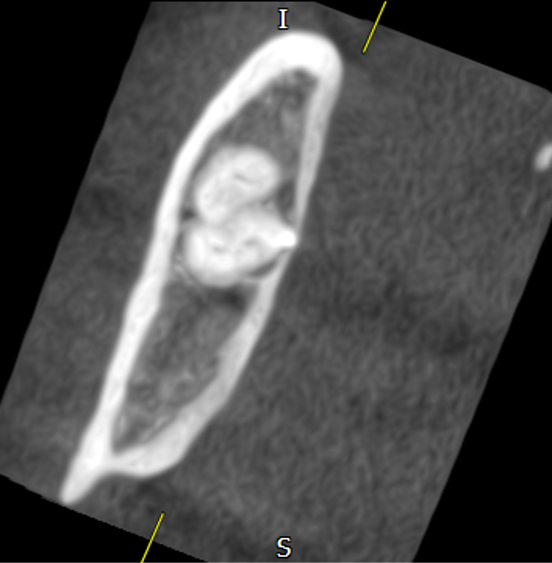
This was taken and reported on by a consultant dental radiologist. This scan showed the LR8 to have three convergent roots and an enamel pearl which protrudes from the mesial root lingually around the middle third of the root (Figures 2, 3, 4 and 5). The inferior dental canal curves lingually around the root deviated and with pronounced narrowing and the enamel pearl lies within this canal and through into the lingual cortication.
The lower left third molar has three fused roots and the inferior dental canal passes compressed between the middle third of the tooth roots and the thinned bony cortex lingually. Both lower third molars keep with an intimate relationship with the inferior dental canal and the roots of the molars. There was no evidence of caries in the lower second molars.
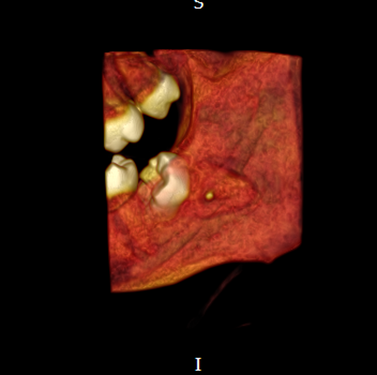
3. Management
Following clinical and radiographic assessment, three management options were considered: retention with no active treatment, coronectomy and complete surgical extraction. The intimate relationship between the mandibular third molar and the inferior dental (ID) canal was an important consideration in treatment planning, given the potential risk of inferior alveolar nerve (IAN) injury during surgical removal. The patient was counselled regarding the risks and benefits of each option, including the potential consequences of both retaining the tooth and undertaking surgical intervention. Current guidance from the Faculty of Dental Surgery of the Royal College of Surgeons of England recommends that treatment planning for mandibular third molars should take into account the current disease status, the likelihood of future disease and the risk of surgical complications, including IAN injury [1].
3.1. Option 1 – No active treatment
The first option was to retain the mandibular third molar with no immediate surgical intervention. This approach would avoid the risk of IAN injury associated with surgical removal of a tooth in close proximity to the ID canal. Where a mandibular third molar is asymptomatic and there is no significant associated pathology, active surveillance may be appropriate, particularly where the risk of surgical complications is considered high [1]. The patient would require appropriate clinical monitoring, with radiographic assessment where indicated, and would be advised to seek review should symptoms develop.
The potential disadvantages of retaining the tooth were also discussed. These include the future development of pericoronitis, dental caries, periodontal disease affecting the third molar or adjacent second molar, root resorption and other pathology such as cystic change [1]. Where pathology is already present, therapeutic removal may be indicated; however, in the absence of significant disease, the risk-benefit balance may favour surveillance when surgical intervention carries a substantial risk of neurological injury [1]. It was also explained that the clinical circumstances may change over time and that extraction could subsequently become necessary if symptoms or pathology developed. In this case, the patient was informed of the increased risk of distal caries in the lower second molars, associated with the angulation of the third molars.
3.2. Option 2 – Coronectomy
The second option considered was coronectomy. This involves removal of the crown of the mandibular third molar while deliberately retaining the roots within the alveolus, with the intention of reducing manipulation of the IAN where the roots are in close proximity to the ID canal. The Faculty of Dental Surgery recognises coronectomy as a technique that may be considered where there is a high risk of IAN injury associated with complete third molar removal [1].
The evidence suggests that coronectomy can significantly reduce the risk of IAN injury compared with complete extraction in appropriately selected high-risk mandibular third molars. In a randomised controlled trial by Renton et al., IAN injury occurred in 19% of patients undergoing extraction compared with no cases following successful coronectomy, although the study included a relatively small number of patients and some intended coronectomies were unsuccessful [2]. More recent systematic reviews and meta-analyses have similarly demonstrated a substantially lower incidence of IAN injury following coronectomy compared with complete extraction [3, 4]. A 2025 systematic review and meta-analysis involving 7,115 third molars reported a relative risk of IAN injury of 0.10 following coronectomy compared with extraction [4].
Despite these potential benefits, coronectomy is not without complications. Retained roots may migrate postoperatively, become exposed or symptomatic, and a small proportion of patients may subsequently require surgical removal [3, 4]. The 2025 meta-analysis reported a surgical reintervention rate of approximately 3.6%, root exposure in 2.7% and a coronectomy failure rate of approximately 2.8% [4]. The patient was therefore informed that coronectomy reduces, but does not completely eliminate, the risk of neurological injury and that long-term clinical and radiographic follow-up may be required. It was also explained that coronectomy may not be appropriate where the roots themselves are associated with pathology or where complete removal is necessary to adequately treat the underlying disease [1].
3.3. Option 3 – Complete surgical extraction
The third option was complete surgical extraction of the mandibular third molar. This would provide definitive removal of the tooth and eliminate the possibility of future pathology arising from the retained third molar or roots. Where a mandibular third molar is associated with significant pathology, including recurrent pericoronitis, unrestorable caries, periodontal disease, resorption, fracture, pulpal or periapical pathology, or associated cystic or neoplastic disease, therapeutic removal may be indicated [1].
However, in this case, the intimate relationship between the roots and the ID canal was recognised as an important risk factor for IAN injury. The patient was counselled that surgical manipulation or mobilisation of the roots could result in temporary or permanent altered sensation in the distribution of the IAN, including the lower lip and chin. The risk of lingual nerve injury, alongside other recognised complications of mandibular third molar surgery such as pain, swelling, infection, bleeding and alveolar osteitis, was also discussed [1].
Overall, the three options were discussed with the patient as part of the consent process. Retention avoided the immediate risk of surgical nerve injury but carried the possibility of future third-molar pathology; coronectomy provided an alternative approach intended to reduce the risk of IAN injury while addressing the crown of the tooth; and complete extraction offered definitive removal but carried a greater potential risk of neurological injury due to the close anatomical relationship with the ID canal. The final management decision was therefore based on the clinical indication for treatment, the radiographic anatomy, the estimated risk of IAN injury and the patient's informed preference. After discussion with the patient they decided they currently only wanted to proceed with clinical monitoring of the lower third molars and surgical removal of the upper third molars under local anaesthetic which was undertaken at a subsequent visit.
4. Discussion
In general, enamel pearls typically occur on maxillary second and third molars, with a prevalence of 1.1–9.7%. [5]. They can have general complications associated with the care of the teeth in question. When more superficial these can increase the amount of food trapping and encourage localised bone loss. In this case the enamel pearl would have limited impact to the cleanability of this tooth. However, it did impact surgical management with the location of the enamel pearl to adjacent structures and therefore impacted on the patient’s surgical management.
Diagnostically, it was not identified on the initial panoramic radiograph, however on review with the CBCT imaging it was significantly more evident. Having reviewed the CBCT imaging it demonstrated anatomical features associated with an increased risk of IAN injury. Using the 3D reconstruction allowed us to highlight the risks to the patient more clearly which allowed her to make an informed decision.
5. Conclusion
In this report we discuss the potential complications associated with an enamel pearl intimately associated with the inferior dental canal on cone beam imaging. In order to reduce potential consequences of inferior dental nerve injury during third molar surgery, the cone beam CT scan and careful consideration of the OPT allow for the patient and surgeon to be well informed about the risk.
References
- Renton T, Coulthard P, Chiu G, Master S, Brookes V, Atack N, et al. Parameters of care for patients undergoing mandibular third molar surgery. London: Faculty of Dental Surgery, Royal College of Surgeons of England; 2020.
- Renton T, Hankins M, Sproate C, McGurk M. A randomised controlled clinical trial to compare the incidence of injury to the inferior alveolar nerve as a result of coronectomy and removal of mandibular third molars. Br J Oral Maxillofac Surg. 2005;43(1):7-12. doi:10.1016/j.bjoms.2004.09.002.[CrossRef] [PubMed]
- Peixoto AO, Bachesk AB, Leal MOCD, Jodas CRP, Machado RA, Teixeira RG. Benefits of coronectomy in lower third molar surgery: a systematic review and meta-analysis. J Oral Maxillofac Surg. 2024;82(1):73-92. doi:10.1016/j.joms.2023.09.024.[CrossRef] [PubMed]
- Kang FW, Yuan XR, Li GC, Yang YFZ, Zhang XM, Hou GY. Coronectomy in lower third molar surgery: a systematic review and meta-analysis. J Oral Maxillofac Surg. 2025;83(5):601-615. doi:10.1016/j.joms.2025.01.014.[CrossRef] [PubMed]
- Moskow BS, Canut PM. Studies on root enamel (2). Enamel pearls. A review of their morphology, localization, nomenclature, occurrence, classification, histogenesis and incidence. J Clin Periodontol. 1990;17(5):275-281. doi:10.1111/j.1600-051X.1990.tb01089.x.[CrossRef] [PubMed]