Back to Article
Video-Assisted Thoracoscopic Surgery (VATS) for Bronchogenic Cyst Resection in a Two-Year-Old Child: A Case Report
Global Journal of Medical Case Reports
| Vol 6, Issue 1
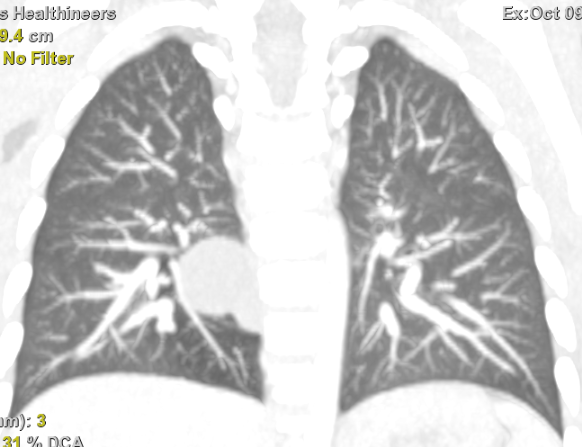
Figure 2
Figure 2.
CT scan - In the superior segment (S6) of the right lower lobe, a paravertebral oval unilocular thin-walled hypodense lesion is identified, measuring approximately 22 × 21 × 20 mm. No definite communication between the lesion and the tracheobronchial tree is demonstrated. In close proximity to this lesion, in the mediastinal paravertebral region posterior to it, an additional small lesion measuring 10 × 8 mm is noted. The latter is abutting the esophageal wall.