
Abstract
Bronchogenic cysts are rare anomalies of the tracheobronchial tree. These are fluid-filled lesions most commonly located in the mediastinum, and less frequently demonstrate communication with the respiratory tract. Diagnosis is typically made prenatally or incidentally on chest radiography during evaluation of respiratory infections. Because of the risk of infection or malignant transformation, bronchogenic cysts require urgent surgical intervention – either excision in cases without communication to the tracheobronchial tree, or pulmonary resection when the cyst is located within the lung parenchyma. We present a clinical case of a two-year-old child who underwent surgical treatment via video assisted thoracoscopic surgery (VATS) for two incidentally diagnosed bronchogenic cysts. Minimally invasive surgical techniques offer several advantages, including substantially decreased postoperative pain, accelerated mobilization and functional recovery, and earlier resumption of normal physical activities.
1. Introduction
Bronchogenic cysts are a rare congenital malformation resulting from abnormal budding of the primitive foregut. The incidence is 1:42.000 to 1:68.000 [1]. They most commonly occur in the mediastinum and are typically unconnected to the tracheobronchial tree, although rare cases of airway communication have been described. These lesions may remain asymptomatic or present with compressive symptoms depending on their size and location In the majority of cases, bronchogenic cysts are diagnosed incidentally during chest radiography performed in the course of a respiratory infection. Clinically, they may also manifest with a mass effect on surrounding tissues or with complications resulting from infection or perforation of the cysts. Malignant degeneration is rare. The definitive treatment is their surgical removal, with minimally invasive surgery increasingly replacing classic thoracotomy.
2. Case report
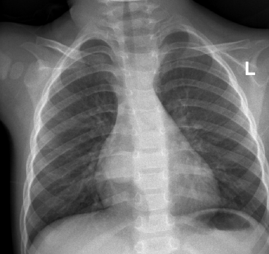
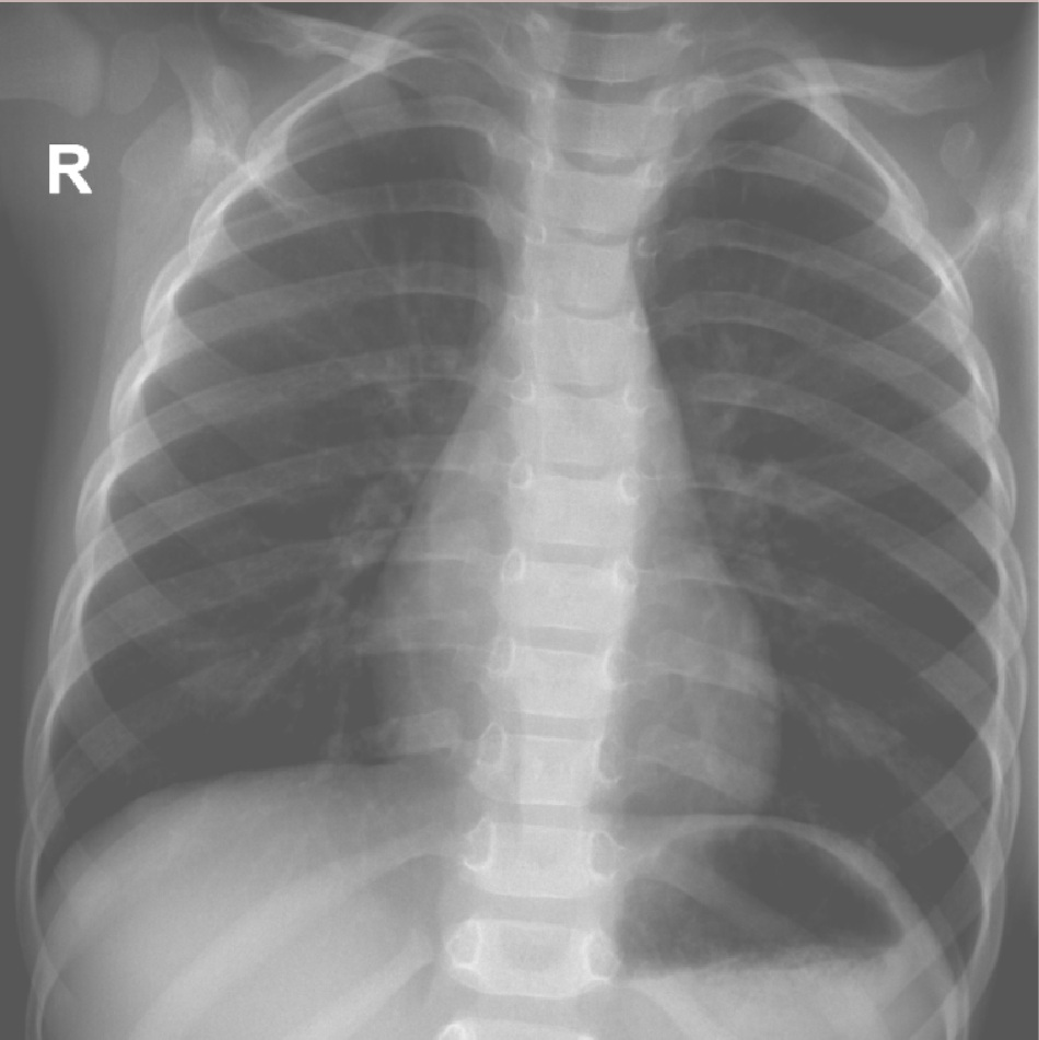
A two-year-old boy with normal development and no significant past medical history presented during an episode of a respiratory infection. A chest radiograph incidentally revealed a cystic formation in the right paracardiac region (Figure 1). Subsequent computed tomography (CT) of the chest demonstrated two well-defined cystic lesions located within the mediastinum. Оne with dimensions 22/20mm located paravertebrally and one smaller with dimensions 10/8mm wall-adjacent to the esophagus (Figure 2 and Figure 3). Upon admission to the pediatric thoracic surgery department, the patient was in good general condition, with normal growth and development for age, and no abnormalities detected on physical examination.
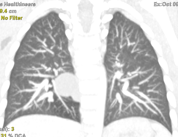
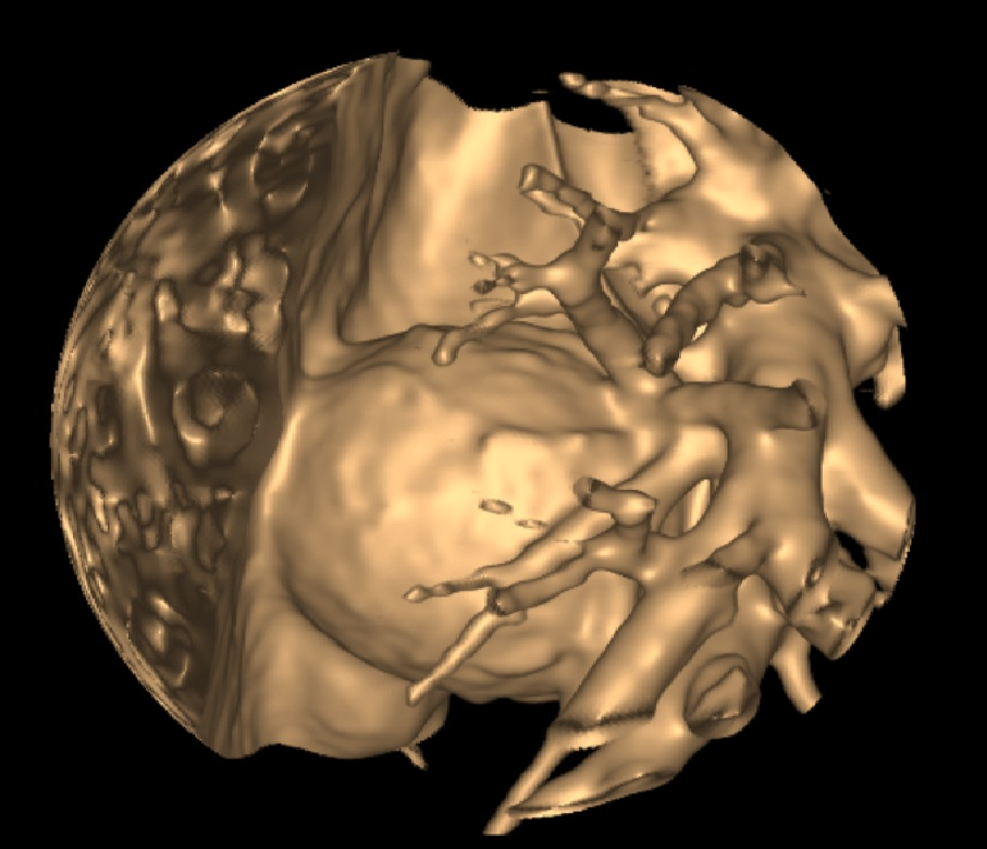
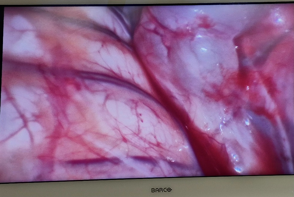
Surgical treatment was undertaken via VATS using four ports under single-lung ventilation. During exploration of the right hemithorax, two cysts were identified adjacent to the inferior pulmonary vein and the esophagus, filled with thick whitish content (Figure 4). The cysts were meticulously dissected with careful hemostasis, and intraoperative evaluation confirmed the absence of any communication with the tracheobronchial tree or esophagus. A single thoracostomy drain was placed for postoperative management.
The postoperative course was uneventful. On the first postoperative day, the nasogastric tube was removed, and the patient was gradually advanced to liquid feeds. The thoracostomy drain was removed on the third postoperative day. The total hospital stay was nine days, and at discharge, the patient was asymptomatic with full enteral intake restored. During follow-up, the child remained clinically well, and a control chest radiograph at one month demonstrated no residual or recurrent lesions (Figure 5). Histopathological analysis confirmed the diagnosis of bronchogenic cysts.
3. Discussion
Bronchogenic cysts are malformations of the tracheobronchial tree that arise from abnormal budding of the foregut during early embryonic development which is not followed by bronchial and vascular development. They were first described in 1859 by Meyer and are among the earliest recognized congenital mediastinal lesions [2]. As a result of abnormal development, a cavity is formed that is most often not connected to the airways and can form at any level of the tracheobronchial tree. Extremely rare, they can be located in the abdominal cavity, neck, skin, pericardium, transdiaphragmatic evolution with an hourglass shape, and retroperitoneum [3]. The formations present as thin-walled cysts with bronchial epithelium, filled with mucus. They may be presented as a single-chamber or multi-chamber formation. Mediastinal cysts typically receive arterial supply from discrete systemic vessels, with venous drainage occurring via the azygos and hemiazygos veins. Due to their usual lack of communication with the tracheobronchial tree, these cysts are rarely subject to secondary infection.
Intrapulmonary bronchogenic cysts are typically thin-walled and usually present as single lesions, although multiple cysts can occur. They most commonly localize to the perihilar regions of the lower lobes. The cysts are supplied by discrete branches of the bronchial arteries, with venous drainage occurring via the pulmonary veins. Unlike mediastinal cysts, they frequently communicate with the bronchial tree, which predisposes them to secondary infection. Such infections can manifest as recurrent pneumonia, fever, sepsis, or other forms of respiratory compromise.
On prenatal ultrasound, bronchogenic cysts typically appear as cystic structures, most commonly within the mediastinum. In some cases, they may cause bronchial obstruction, leading to distal airway dilation and overdistension of the affected lung.
Postnatally, the majority of patients with bronchogenic cysts remain asymptomatic. Clinical manifestations typically arise as the cyst enlarges, causing compression of adjacent structures such as the lung parenchyma, trachea, esophagus, heart, great vessels, or even intra-abdominal organs. Potential complications include infection of the cyst, rupture leading to hemothorax or pneumothorax, and, rarely, malignant transformation [4]. Documented malignancies arising from bronchogenic cysts include rhabdomyosarcoma, pleuropulmonary blastoma, anaplastic carcinoma, leiomyosarcoma, and adenocarcinoma, although such events are exceedingly uncommon.
In the differential diagnosis, congenital pulmonary airway malformation (CPAM), mediastinal or intrapulmonary tumors, foregut duplications, and diaphragmatic hernia should be considered.
On conventional radiography, uncomplicated bronchogenic cysts typically appear as well-circumscribed, rounded opacities of soft tissue density. Compression of adjacent structures may occasionally be observed. In some cases, calcium oxalate deposits within the cyst can produce a radiopaque focus at its base, known as “calcium milk.” In complicated cysts, imaging may reveal blurred or thickened cyst walls, the presence of an air-fluid level, or an entirely air-filled cavity [5].
Contrast-enhanced CT or magnetic resonance imaging confirm the malformation and its relationship to the adjacent structures. The cysts present as a well-defined or spherical mass with varying intensity.
All bronchogenic cysts are subject to surgical treatment due to the risk of complications. The timing of intervention depends on the clinical presentation. In symptomatic newborns, emergency surgical treatment may be required in cases of significant compression on adjacent structures affecting respiration and/ or hemodynamics. For prenatally detected bronchogenic cysts that are asymptomatic, surgical intervention is elective and is typically performed within the first year of life. As with other pulmonary malformations, some surgeons proceed to earlier surgical treatment. In cases where the bronchogenic cyst is an incidental finding, surgical intervention is elective, within a short time after diagnosis due to the risk of complications. Surgical treatment consists of excision of the cyst, and in cases of intrapulmonary location – pulmonary resection of the appropriate extent.
4. Conclusion
This case highlights the importance of early recognition and timely surgical management of bronchogenic cysts, even when incidentally detected, in the pediatric population. These rare congenital malformations, while often asymptomatic, carry the potential for significant complications—including infection, compression of mediastinal structures, and, albeit rarely, malignant transformation. Surgical excision remains the definitive treatment, with minimally invasive approaches such as video-assisted thoracoscopic surgery offering the dual benefit of complete lesion removal and minimal physiological disruption [6].
Beyond the technical aspects, this case underscores a broader principle in pediatric thoracic care: the need to balance vigilance with intervention. Even in clinically silent cases, the unpredictable natural history of bronchogenic cysts justifies a proactive approach, allowing preservation of pulmonary function and prevention of life-threatening sequelae. Moreover, the anatomical variability and potential for airway or vascular involvement demand precise preoperative imaging and meticulous surgical planning, emphasizing that individualized, anatomy-driven management is central to achieving optimal outcomes.
Ultimately, this case exemplifies how careful diagnostic evaluation, interdisciplinary planning, and minimally invasive surgical techniques can transform the management of a rare congenital anomaly from a theoretical risk into a predictable, safe, and curative outcome, reinforcing the paradigm of organ-preserving, patient-centered pediatric thoracic surgery.
References
- Gross DJ, Briski LM, Wherley EM, Nguyen DM. Bronchogenic cysts: a narrative review. Mediastinum. 2023 Apr 20;7:26. doi: 10.21037/med-22-46. PMID: 37701638; PMCID: PMC10493618.[CrossRef] [PubMed]
- Meyer, H. Ueber angeborene blasige Missbildung der Lungen, nebst einigen Bemerkungen über Cyanose aus Lungenleiden. Archiv f. pathol. Anat. 16, 78–95 (1859). https://doi.org/10.1007/BF01945249[CrossRef]
- Reichelt O, Grieser T, Wunderlich H, Möller A, Schubert J. Bronchogenic cyst. A rare differential diagnosis of retroperitoneal tumors. Urol Int. 2000;64(4):216-9. doi: 10.1159/000030534. PMID: 10895088.[CrossRef] [PubMed]
- Whooley J, White A, Soo A. Bronchogenic cyst: a rare case of malignant transformation. BMJ Case Rep. 2022 Apr 4;15(4):e248916. doi: 10.1136/bcr-2022-248916. PMID: 35379683; PMCID: PMC8981341.[CrossRef] [PubMed]
- Jones J, Le L, Silverstone L, et al. Bronchogenic cyst. Reference article, Radiopaedia.org (Accessed on 20 Jan 2026) https://doi.org/10.53347/rID-8374[CrossRef]
- Adams S, Jobson M, Sangnawakij P, Heetun A, Thaventhiran A, Johal N, Böhning D, Stanton MP. Does thoracoscopy have advantages over open surgery for asymptomatic congenital lung malformations? An analysis of 1626 resections. J Pediatr Surg. 2017 Feb;52(2):247-251. doi: 10.1016/j.jpedsurg.2016.11.014. Epub 2016 Nov 13. PMID: 27889066.[CrossRef] [PubMed]